Medical Illustration

Medical Illustration

Fred Harwin is a Medical Illustrator. He received his Master of Science degree in medical and biological illustration from the University of Michigan. After completing school, Harwin served as the Director of Medical Illustration at Wayne County General Hospital, a teaching affiliate of the University of Michigan.

Medical Illustration by Fred Harwin

He later joined the faculty at Oregon Health Sciences University in Portland, Ore. Harwin has also taught at Portland State University, the Oregon Museum of Science and Industry and was an Adjunct Professor of Medical Illustration at the University of California's San Francisco Medical Center.

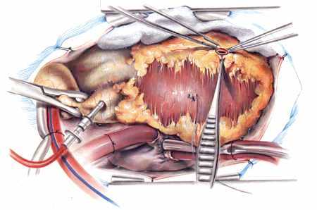
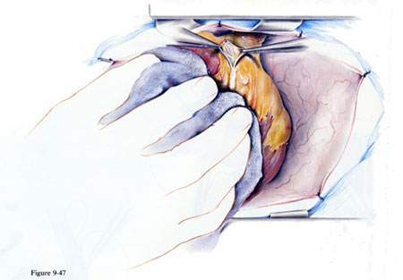
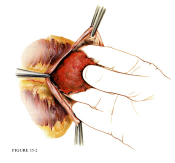
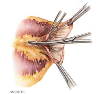
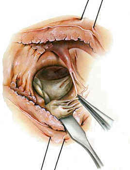
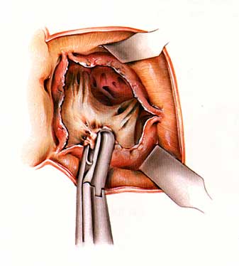
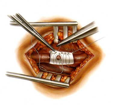
Among Harwin's favorite creations are Volumes I and II of the Manual of Cardiac Surgery. Both are full-color primers of instructional illustrations and text depicting cardiac operating procedures from the surgeon's perspective. Listed on the manuals' covers in unprecedented fashion as co-author with cardiac surgeons Bradley J. Harland and Albert Starr, Harwin labored 3 1/2 years on some 310 color illustrations.

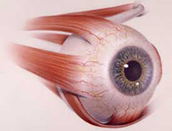
The images below provide an example of Fred Harwin's work from surgical to anatomical illustrations.

surgery 1

Distal Left Anterior Descending Artery Anastomosis

The heart is placed on pads to elevate the anterior wall and bring the left anterior descending coronary artery medially. The epicardium over the artery and the artery are incised

surgery 2

Distal Obtuse Marginal Artery Anastomosis

The heart is retracted by hand. with the apex toward the patient's right shoulder. The epicardium is incised, exposing the artery.

surgery 4

Left Ventricular Aneurysm

Intramural clot is carefully scooped out.

surgery 3

Left Ventricular Aneurysm

The middle of the aneurysm is incises and the incision is extended with scissors.

surgery 5

Tetralogy of Fallot

Incision and excision of the septal and parietal bands help expose the ventricular septal defect. Exposure is further aided by placing a small intracardiac sucker through the defect, with retraction to the left.

surgery 7

Aortic Valve Surgery

Seen in this illustration is a "plane" that can usually be located between the calcium and the leaflet and the calcium is peeled off

surgery 8

Goretex Patch

Goretext patch surgical procedure.

surgery 6

Endoscopic Surgery

Anastomosis using robotics.